This assignment gives you the opportunity to demonstrate your ability to use the basic functions of Deep View. You will study a protein, which will also be the protein on which you will write your enzyme paper later in the semester. You will prepare some specified views of your protein, save them as Deep View coordinate files, and turn them in the files for evaluation. Finally, you will write a brief description of the protein, some of its structural features, and its interaction with its hetero group. Note that you will also use at least some of these images in your enzyme paper later in the semester!
Select a protein from the list here to use for the assignment. Each student will do a different protein. Email your selection to Dr. Osborne for approval. All the proteins contain a cofactor or inhibitor. If you prefer, you may choose and study a different protein not on the list and of particular interest to you, but it must function as a single chain and contain a hetero group. In addition, it must be a protein that no one else in the class has already chosen. Proteins with more than 250 residues can be very difficult for graphics beginners, so smaller is better. Email your selection to Dr. Osborne for approval.
Campus computer lab computers have Deep View (also known as the Swiss-PDBViewer) installed, already. Go to Windows (Start) menu and open the [MU Applications] folder. If you want to install this software on your personal computer computer, go ahead and download it.
Whether you accept an assigned protein or find your own, obtain your file from the Protein Data Bank using a web browser by saving it to the desktop or else import it directly to DeepView from the PDB server using File: Import in Deep View .
Download the 1HEW.pdf file from Canvas to then open from Deep View for use in the tutorial. Go through the Deep View tutorial Sections 1 through 6. Then, use what you learn to create the views described in the following list. Save each view with a filename in this format: xxxx#.pdb, where xxxx denotes the PDB file code for your protein, and # is a one-digit number (example: 1hew3.pdb).
Once you have constructed the required view save it as follows:
File: Save: Layer...
The familar file-saver dialog box appears. Navigate to the
location where you want Deep View to store the file, type in the
appropriate file name, and click Save. At the end of this
exercise, you will combine all of your files into one Deep View
Project File (instructions below).
If your protein contains two or more identical chains, restrict all views to one chain and its associated hetero group. If there are hetero groups in addition to the one listed in the table of assigned files, include only the hetero group listed in the table. Finally, do not leave water molecules on display in any of your completed views. You may find it convenient to create a working file containing only the one chain that you plan to study, with its hetero group. The command File: Save: Selected Residues Only... is useful for this purpose.
The command File: Save in Original Orientation is no longer active.
Deep View saves coordinate files in their original orientation if you use any of these commands:
Deep View saves coordinate files in their current orientation if you use this command:
Following is the list of views to make and save. Each is accompanied by an example -- a convergent stereo snapshop of cytochrome b5 (PDB 3b5c) in the assigned view. Cytochrome b5 is a liver redox protein that contains a heme prosthetic group.
Although most of the views below are shown using a wire frame style, you might prefer to render all the views in a more realistic 3D view by changing the following two settings: Display: Use OpenGL Rendering and then Display: Render in solid 3D. As a result, your views will all be nicer than those shown stereo, wire frame images below because yours will not be in wire frame style. You can play with the appearance of rendered views by adjusting things in Preferences: 3D Rendering and Preferences: 3D Lights. Press <Ctrl>-3 will toggle between this 3D and normal viewing modes. The OpenGL Rendering in solid 3D model of cytochrome b5 looks like this (after the inevitable degradation of conversion to a GIF file):
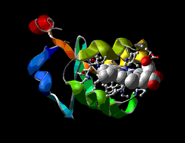
View 1 (filename xxxx1.pdb): Protein in ribbon model only, colored by secondary structure. Ligand (hetero group) colored CPK with dotted van der Waals surface. Note that van der Waals surface is the volume effectively occupied by an individual atom or molecule.
Example:
View 2 (filename xxxx2.pdb): Detail of an alpha helix (if your protein contains no alpha helix, you can skip to View 4). Mainchain colored CPK, side chains colored by type. Compute and display hydrogen bonds, and arrange the view to emphasize hydrogen bonding (Hbonds will not be saved). Label the terminal residues of the helix.
NOTE: Due to a bug in SPV 3.5, this file may be saved with both mainchain and sidechain colored by type.
For your written description, note the distribution of polar and nonpolar residues around the helix.
Example:
Note that most of the residues pointing to the right and back are polar. Three of the residues pointing left are nonpolar (gray).
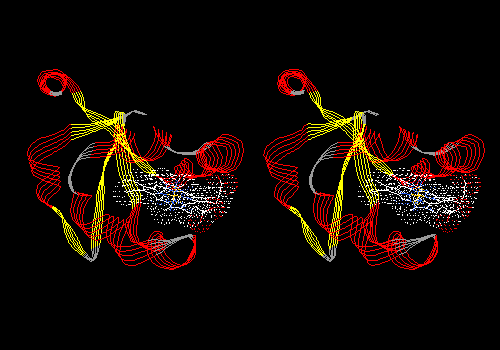
View 3 (filename xxxx3.pdb): Same helix as in View 2 in the context of entire protein. Remainder of protein in ribbon model from View 1. No ligand. No labels. Be sure that ribbon is NOT shown for residues in the helix under examination (Select: Inverse Selection may be a handy command in making the ribbon part of this view).
For your written description, note the location (surface, buried) of the helix. In the description, relate the location to the distribution of residue types as shown in View 2.
Example:
Note that the helix is on the surface, with much of its surface exposed to solvent. Thus most of its sidechains are polar. The three nonpolar side chains are on the interior of the helix. In later views, you can see that these side chains are in contact with the heme prosthetic group.
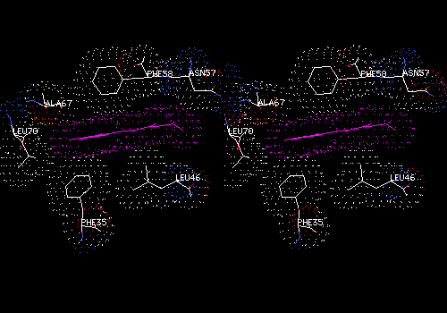
View 4 (filename xxxx4.pdb): Detail of 3 or 4 strands of pleated sheet (if your protein contains no pleated sheet, skip to View 6). Mainchain CPK, side chains by type. Labels on termini of each strand (might not save). Arrange view to emphasize hydrogen bonding (H-bonds not saved).
For your written description, note the distribution of polar and nonpolar residues on opposite sides of sheet.
Example:
Note that both sides of this three-strand sheet contain mostly nonpolar side chains (gray -- tyr is yellow, but is also mostly nonpolar). Some residues at the top and bottom edges are polar. The next view shows why.
View 5 (filename xxxx5.pdb): Same strands as above in contex of entire protein. Remainder of protein in ribbon model as in View 1. No ligand (hetero group). No labels. No hydrogen bonds. Be sure that ribbon is NOT shown for residues in the sheet under examination.
For your written description, note the location (surface, buried) of the sheet. Relate the location to the distribution of residue types on opposite sides of the sheet.
Example:
Note that the side of the sheet facing away from the viewer is buried and contains mostly nonpolar (gray) sidechains. Surprisingly, several side chains on the nearer, exposed face of the sheet are also nonpolar. This form of cytochrome b5 is actually a fragment cut from a larger protein. In the intact protein, most of this sheet if buried, and thus most side chains on both sides of the sheet are nonpolar. The sheet forms the back of a pocket that holds the heme.
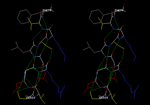
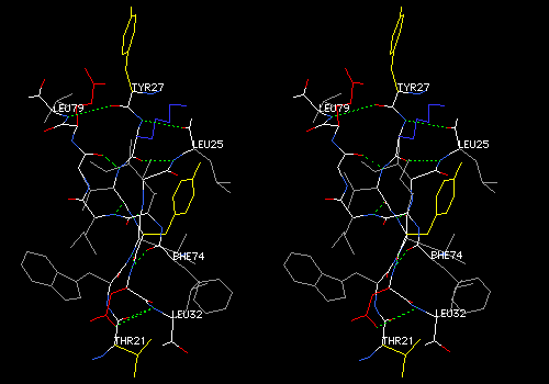
View 6 (filename xxxx6.pdb): Ligand (hetero group) with residues that lie within 5.0 angstroms of the ligand. Mainchain and ligand colored CPK, side chains colored by type. Ligand labeled. Compute H-bonds and arrange the view to emphasize hydrogen bonds between ligands and protein.
For your written description, make a list of hydrogen bonds that link the hetero group to the protein. For each bond, list donor and acceptor atoms, using PDB atom designations the Deep View uses in its labels. If any hydrogen bonds from or within the hetero group appear to be erroneous, remove them (Build: Remove H-Bond...), and mention them in your description.
CRITERIA FOR IDENTIFYING HYDROGEN BONDS AND SALT LINKS: Remember that hydrogen bonds involve two electronegative atoms that share one hydrogen atom. In proteins, hydrogen bonds involve mostly N and O, but never C. A thiol sulfur of cysteine can form a weak hydrogen bond with N or O, but not with another S. Hydrogen atoms are not visible in electron density maps from protein crystallography, so they are not shown in protein models. Therefore, we must infer the presence of a hydrogen bond when we find a pair of appropriate atoms within about 2.8 angstroms of each other. In addition to chemical criteria (one atom must be of a type that is normally protonated, one atom must be N or O, and the other must be N, O, or S) and distance criteria (2.8 ± 0.5 angstroms), the atoms must satisfy geometric criteria for hydrogen bonding. The hydrogen is involved with a bonding orbital on one atom, and with a nonbonding orbital on the other, so a line extending from one hydrogen-bonded atom to another must make either a tetrahedral or trigonal angle with the other covalent bonds at both atoms. Salt bridges, or electrostatic interactions, must involve atoms of opposite charge. The atoms can be in almost any orientation to each other, at distances near the van der Waals contact distance of about 3 Å.
Example:
In the foreground, a heme carboxyl makes two hydrogen bonds with a serine reside (yellow side chain). One bond is to the main-chain NH, and the other to the side-chain OH of serine. Deep View draws some erroneous hydrogen bonds between the histidine ring about the heme iron to the heme nitrogens. These bonds have been removed. All this is hard to see without stereo.
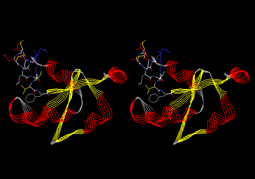
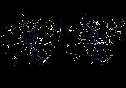
View 7 (filename xxxx7.pdb): Same as View 6, but van der Waals surface dots on all atoms. Ligand (hetero group) colored a bright shade of purple (brighter than the example below). Ligand surface colored CPK (although the image below shows ligand surface as purple, use CPK, which is the default). Mainchain AND sidechains colored CPK. Explore this view by slabbing (Display: Slab). Adjust slab depth to 5 angstroms in the Display Window Attributes dialog. Hold down <Ctrl> while translating to move the slab forward and back. Find and save a view in which some specific ligand-protein contacts are evident. The ligand will probably be cut in cross section in such a view. Add some labels to show which residues are involved in contacts.
Example:
This cross-section of the nonpolar heme group (purple) shows surface contacts with several nonpolar sidechains.
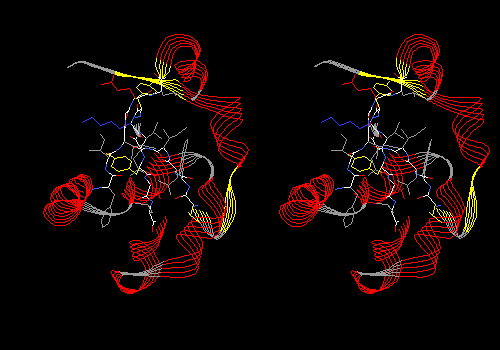
View 8 (filename xxxx8.pdb): Ribbon display of mainchain, colored by secondary structure succession. Ligand (hetero group) in CPK with dotted van der Waals surface. Side chains (but no main chain) within 5.0 angstroms of ligand in CPK. This is similar to the first view of a protein you might see in a textbook.
NOTE: Due to a bug in SPV 3.5, the setting Display: Show Sidechains even when Backbone is hidden is not saved. In written description, instruct the reader to turn on this function.
Example:
NOTE: Save pictures that have been rendered in solid 3D for use in reports and web pages with the command File: Save: Image, which saves a .png file. The .png picture can then be directly inserted into a Word or PowerPoint document.
Test-run each of your files by opening them in Deep View. Open them in the order created, and do not close each file before opening the next. You will have all eight files loaded at once.
Wind: Layer Infos
This command opens the Layer Information window, which
allows you to manage multiple files. Each file that you load becomes
a separate layer.
<ctrl>-<tab>, <tab>,
<tab>, ... (hold down the ctrl key, and press tab
repeatedly.
This operation is called "cycling" or "blinking". Each time you press
tab (while holding down ctrl), you turn off display of
the currently displayed layer, and turn on display of the next layer
on the list. Thus you can move easily from one of your views to the
next. Blink through all your views and make sure that each has the
correct settings (review instructions, above, for each view). Correct
settings as needed. As you make these changes, you are not changing
any of your saved files.
When all views are as you want them, arrange View 1 into a photogenic pose. Next you will save the entire project, and Deep View will save all files in their current orientation.
Save: Project
Name the project file "#xxxProj.pdb", and save it to your
disk or a convenient location. Quit Deep View, start it again, and
open your project file. Do a final check of all views to be sure they
are as you want them.
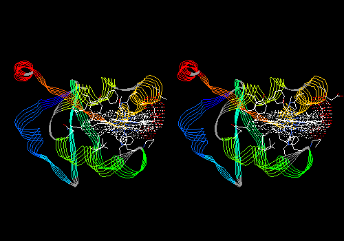
![]()
To complete this assignment, write a brief description and structural analysis of your protein, illustrating it with the views saved in your project file, and presenting the information you were asked to gather as you made each view. Assume that your reader knows how to use Deep View, and that your reader will open your project file and move from view to view as you discuss your protein. Assume that your reader's knowledge of protein structure is similar to yours.
Give the description an informative title, and list yourself as the author.
In the first paragraph, give the name and source (organism) of the protein, and then describe its function and the function of its hetero group. If it is an enzyme, give the reaction that it promotes, including molecular structures of substrate(s) and product(s) that you draw yourself. ChemSketch is an excellent, free program for drawing molecular structures in a word-processor document. It is available for free on the web and installed on all campus computers. References used must be cited in the text.
In the second paragraph, describe the structure, including 1) the number of chains and number of residues in each, 2) major secondary structural elements and the residue numbers that they encompass, and 3) the location among your structural elements of the hetero-group binding site. Insert into the document the .png image files of the appropriate views. Note that you export these .png image files by doing File/Save/Image.
In the third paragraph, describe the binding of the hetero group to the protein. Insert into the document the .png image files of the appropriate views. Include description of the protein-hetero H-bonds, as well as hydrophobic and other types of interactions. Be sure to look carefully at all groups within 5 angstroms of the hetero group so that you do not miss important interactions.
Model your written description after the introductory description of any protein (for example myoglobin) in a biochemistry textbook. Write this description with a word processor, print it, and save it to the same location as your project file. Submit your file in one of the following formats: Microsoft Word, OpenOffice, or RTF.
Here are some potential sources of information about your protein and its hetero group, which you will cite throughout the text:
![]()
When you have completed your project file and description, you are ready to submit them to me, upload the description and structural analysis Word document and the Swiss-PdbViewer project file(s) to the dropbox on Canvas. Also, turn in a hard copy of your description and structural analysis.
This assignment counts as 40 points.
![]()
CHEM-405 Biochemistry I
Fall 2017
Email the name of your chosen protein to Dr. Osborne for approval.
|
|
Enzyme |
|
|
Phthalate dioxygenase reductase |
FMN |
||
| Hannah Janson | Galactose-1-phosphate uridyltransferase |
Fe3+ |
|
| Adam Newport |
Adenylate kinase |
ATP-analog inhibitor AP5A in two conformations |
|
|
DNA (cytosine-C5-)-methyl transferase |
S-adenosyl-L-methionine |
||
| Maeleigh Tidd | human cytochrome P450 3A4 |
heme | |
|
Transketolase (2 chains -- use one) |
thiamine pyrophosphate |
||
| 1X8R | 5-Enolpyruvylshikimate-3-phosphate synthase | (S)-phosphonate analogue | |
MacKenzie VanCoutren |
Human udp-glucuronosyltransferase 1-1 |
selenomethionine |
|
| Anthea Ayebaze |
Human carbonyl reductase |
NADP |
|
| Kyra Zadylak |
Methionine synthase (2 chains - use one) |
Vitamin B12 |
|
| Yafet Leake |
Dihydrofolate reductase (2 chains -- use one) |
folic acid |
|
| Nicky Patel |
Acetyl-CoA carboxylase |
biotin |
|
| Joel Brenneman |
HIV-1 protease (2 chains -- use both) |
sulfamide inhibitor AHA006 |
|
| Courtney Hersick |
Metallo-beta-lactamase |
Fe3+ |
|
| William Mikesell |
Human cathepsin D |
N-(3,4-dimethoxybenzyl)-Nalpha-{N-[(3,4-dimethoxyphenyl)acetyl]carbamimidoyl}-D-phenylalaninamide |
|
| Peyton Banks |
Glutathione reductase |
FAD |
|
| 1Q21 | Ras protein catalytic domain | GDP | |
| Trish Cook | 4C5P |
arylamine n-acetyltransferase |
azide |
| Emily Centofanti | 2F92 | Farnesyl diphosphate synthase | alendronate |
| Rachael Samm | 4CEX | Urease |
Ni2+ |
| Samuel Tetteh-Quarshie | 2AIA | Peptide deformylase | Sb-543668, a potential pneumonia drug (2 superimposed models in alternative conformations) |
| Judy Truong | 1S8G | Phospholipase A2 | dodecanoic acid |
| Ryan DeMars | 5FPQ | Human acetylcholinesterase | O-[(S)-methyl(1-methylenethoxy)phosphoryl]-L-serine |
| Andy Giles | 1HPC | H-protein of glycine cleavage system | lipoic acid |
| Nathan Wilkinson | 1ALL | Allophycocyanin (2 chains -- use one) | phycocyanobilin |
| Brittany Koehl | 1PAH | Human phenylalanine hydroxylase dimer |
Fe3+ |
| Brock Spangle | 3BLP | Human salivary alphy-amylase | Ca2+ |
| Joshua Madueke | 1GPE | Glucose oxidase | FAD |
| Nicole Osborne | 1PRH | Prostaglandin H2 synthase-1 | heme |
| Olivia Janson | 1HE3 | Biliverdin IX Reductase | NADP and biliverdin (analyze binding to either one) |
| Cole Wolf | 4AB1 | Human carboxylesterase 1 |
N-acetyl-D-glucosamine |
| Kitt Hersick | 1LHC | Alpha-thrombin | peptide-analog inhibitor |
| Ashlyn Teders | 2VVB | Carbonic anhydrase II (human) | Zn2+ | Sara Carder | 2GK1 | beta-Hexosaminidase A (H. sapiens) | NAG-thiazoline in active site | Olivia Jenks | 1G8F | ATP Sulfurylase (S. cerevisiae) | SO42- in active site |
| Jacob Ahlgrain | 1UWS | Beta-glycosidase (lactase) | 2-Deoxy-2-fluoro-glucose |
| Zach Martin | 4WNC | Glyceraldehyde-3-phosphate dehydrogenase | NAD |
| Sherry Wong | 4QOL | Catalase | heme |
| Holly Conway | 6ADH | Alcohol dehydrogenase (horse) | nicotinamide adenine dinucleotide |
| Haleigh Fernandez | 4DM9 | Ubiquitin Carboxy-terminal hydrolase L1 | fluoromethyl ketone Z-VAE(OMe)-FMK |
![]()